A 14-Day Recovery and Physical Activity Levels After an Ankle Sprain in Mice.
Hubbard-Turner T, Wikstrom EA, Turner MJ.J Athl Train. 2019 July
Full text freely available
Prolonged Rest, Long-Term Dynamic Balance, and Gait in a Mouse Ankle-Sprain Model.
Wikstrom EA, Hubbard-Turner T, Duncan A, Cline J, Turner MJ. J Athl Train. 2019 July
Full text freely available
Take Home Message: Longer rest and recovery periods following an ankle sprain may lead to better short- and long-term physical activity and dynamic balance.
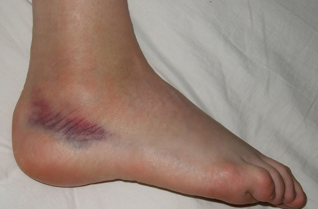
While an ankle sprain is the most common athletic injury, many people think of it as a harmless injury with a swift recovery. However, increasing evidence suggests that a majority of people after an ankle sprain will have long-term disabilities such as chronic ankle instability, reduced physical activity, and worsened health-related quality of life. Therefore, the research group used a mouse-model to identify short- and long-term effects of different lengths of rest and recovery after a surgically induced ankle sprain of the talofibular and calcaneal fibular ligaments. The authors randomly assigned 18 male mice to one of three groups following surgery: 3 days, 7 days, or 14 days of rest. During rest periods, the mice were free to ambulate in their cages but refrained from voluntary physical activity. Following the rest period, a running wheel with magnetic sensors and digital odometers were placed inside the cages for the mice to voluntarily begin exercising. The investigators inspected daily the odometers and recorded running duration (minutes) and distance (kilometers). They calculated average daily speeds (m/min) for each week. Further, the mice were trained to performed dynamic balance and gait assessments before (baseline) and at 3 days, 1, 2, 4, 6, 12, 18, 24, 36, 42, and 48 weeks after injury. Dynamic balance was assessed using a 1 meter long, round wooden beam on a 15º incline. Mice were allowed 60 seconds to cross the beam and the average of 2 trials were taken, along with the amount of times sprained ankle limb slipped off the beam. Gait was assessed using a footprint test, which measured stride length of the sprained limb. Hubbard-Turner and colleagues found that all 3 groups had similar exercise duration, distance, and speed from 3 to 15 weeks post-surgery. However, the 14-day rest group had greater exercise distance and speed during the 16 to 45 week follow-up period and also faster speed starting at 31 weeks after surgery compared to the 3- and 7-day rest groups. Similarly, Wikstrom and colleagues revealed the mice in the 14-day rest group had better dynamic balance and crossed the beam faster than mice in the in 3-day rest period.
The overall conclusion from these two articles highlight the importance of adequate rest following an acute ankle sprain. While there was little evidence of prolonged rest benefiting short-term recovery, the 14-day rest group had a better long-term recovery of physical activity and balance compared to the shorter rest groups. While the authors acknowledged the time frames used in this mouse-model fail to directly translate to human-time, we can make inferences into the utility of these results. For instance, 70% of athletes will return to sport three days after an acute ankle sprain and 90% will return to activity after 1 week. Moreover, more than 50% of those who sprain their ankle report residual symptoms for more than 10 years. We also know that between 40 to 70% of individuals who sprain their ankle will develop chronic ankle instability, which is associated with poor balance, muscle weakness, abnormal gait patterns, and a reduced health-related quality of life. Furthermore, when taking into consideration the inflammatory process following an acute injury, physiological evidence would suggest tissue recovery needs 6 to 12 weeks to restore adequate tensile strength. As healthcare professionals treating athletes, our goal is to facilitate a safe return to play after an acute injury. However, we need to start defining “safe” return to play as not just short-term effects but also the long-term effects. Allowing our patients to return to play as quickly as possible following an injury may not yield the best long-term results. In a community often focused on today, we need to be advocates for the patient’s long-term wellness.
Questions for Discussion: What is the recommended rest time you suggest to your patients following acute ankle sprains? Do you notice a relationship between longer rest and better recovery from ankle sprains?
Written by: Danielle M. Torp
Reviewed by: Jeffrey Driban
Related Posts:
Mid-Life Ankle Crisis with Chronic Ankle Instability
High School Ankle Sprain “Care”cteristics
Acute Care Needs to Focus More on Preventing Chronic Ankle Issues

There is large heterogeneity of neuromusculoskeletal impairment following lateral ankle sprains (LAS). This is likely a variability of injury mechanism, anatomic injury, and patient phenotype. This is also likely contributory to why there are is such heterogeneity of clinical findings in patients who develop Chronic Ankle Instability (CAI). [1]
Tendon injuries, such as tears of the fibularis longus, fibularis brevis, extrinsic flexors and extensors, or their retinaculum are present in 12%[2] to 30%[3] of patients following LAS. Bony contusions are common following inversion injury. Talar contusions are present in 44%[2] to 50%[3] of all inversion injuries and 76% of lateral ligament injuries.[3] Tibial and fibula contusions occur in 23% and 12% of inversion sprains, respectively, and calcaneal and navicular contusions in 8% of the cases.[2] Occult fractures are also fairly common following inversion sprain (14%[4] to 22%[3]), with 5th metatarsal fractures[4] and lateral malleolar avulsions[3] being the most common. In addition to osseous involvement, both tibial and fibular nerves are often injured following lateral ankle sprain. Decreased nerve conduction velocities and increased latencies in patients that have incurred lateral ankle sprain have been reported for the fibular nerves (17% of patients with a grade 2 injury; 86% of patients with a grade 3 injury) and tibial nerve (10% of patients with a grade 2 injury; 83% of patients with a grade 3 injury).[5] Similar findings have been found in the tibial and fibular nerves in soccer players with lateral ankle sprain or chronic ankle instability.[6] Patients who go on to develop CAI also have persistent decreased nerve conduction velocities in the superficial fibular nerve.[7]
Due the wide heterogeneity of injury patterns following LAS, it is imperative for clinicians to perform a comprehensive evaluation. The period of protection and optimal loading will need to be tailored to the individualized patient and dependent on the evaluation and our knowledge of tissue healing.
[1] Hertel J, Corbett RO. An Updated Model of Chronic Ankle Instability. J. Athl. Train. 2019;54:572–588.
[2] Roemer FW, Jomaah N, Niu J, et al. Ligamentous injuries and the risk of associated tissue damage in acute ankle sprains in athletes: a cross-sectional MRI study. Am. J. Sports Med. 2014;42:1549–1557.
[3] Khor YP, Tan KJ. The anatomic pattern of injuries in acute inversion ankle sprains. Orthop. J. Sports Med. [Internet]. 2013 [cited 2018 Jul 10];1. Available from: https://www.ncbi.nlm.nih.gov/pmc/articles/PMC4555519/.
[4] Fallat L, Grimm DJ, Saracco JA. Sprained ankle syndrome: Prevalence and analysis of 639 acute injuries. J. Foot Ankle Surg. 1998;37:280–285.
[5] Nitz AJ, Dobner JJ, Kersey D. Nerve injury and grades II and III ankle sprains. Am. J. Sports Med. 1985;13:177–182.
[6] Jazayeri Shooshtari SM, Didehdar D, Moghtaderi Esfahani AR. Tibial and peroneal nerve conduction studies in ankle sprain. Electromyogr. Clin. Neurophysiol. 2007;47:301–304.
[7] Simon J, Docherty C. Slower nerve conduction velocity in individuals with functional ankle instability. Int. J. Sports Med. 2014;35:731–736.
Hello John,
Thank you for the informative review of common, yet hetergeneous, impairments following inversion sprains and ankle injuries.
I could not agree more with your perspective of clinicians needing to implement immobilization or optimal loading strategies on an individualized patient level. I believe the utility of research studies, as the one in this post, are useful in helping clinicians understand the importance of re-introducing physical activity in a safe manner in coordidance with appropriate tissue healing. I think far too often the acute signs and symptoms fade quickly, leaving the patient feeling much better, yet the tissues and structures are not physiologically ready to withstand loads introduced by physical activity. Clinicians should be aware of this discrepency and upon their assessment of patient readiness make an evidence-based decision regarding loading strategies during the rehabilitation process.